















Spine Plus Blog
Bulging Disc Treatment
Published: 19 February 2015 · 38-procedure reference guide
Overview
Intervertebral discs function as cushions between spinal bones and protect the nerve roots that pass between them. Problems develop through injury or degenerative changes and fall into three main categories.
Loss of Disc Height
Spondylosis
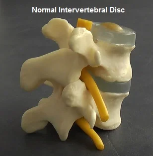
Discs thin progressively, increasing pressure on facet joints and potentially compressing spinal nerves as the disc space narrows.
Torn Disc
Annular Tear
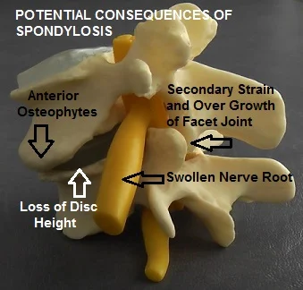
The outer wall of the disc ruptures due to trauma or poor core stabilisation. This can be acutely painful, even without nerve root involvement.
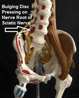
Bulging Disc
Herniation or Prolapse
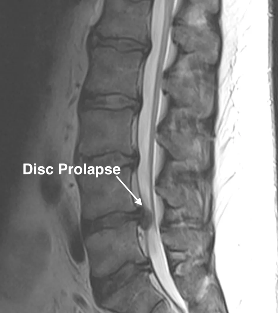
An outpouching of the normal contour of a disc can exert pressure and irritation on the neighbouring spinal nerve to cause sciatic pain.
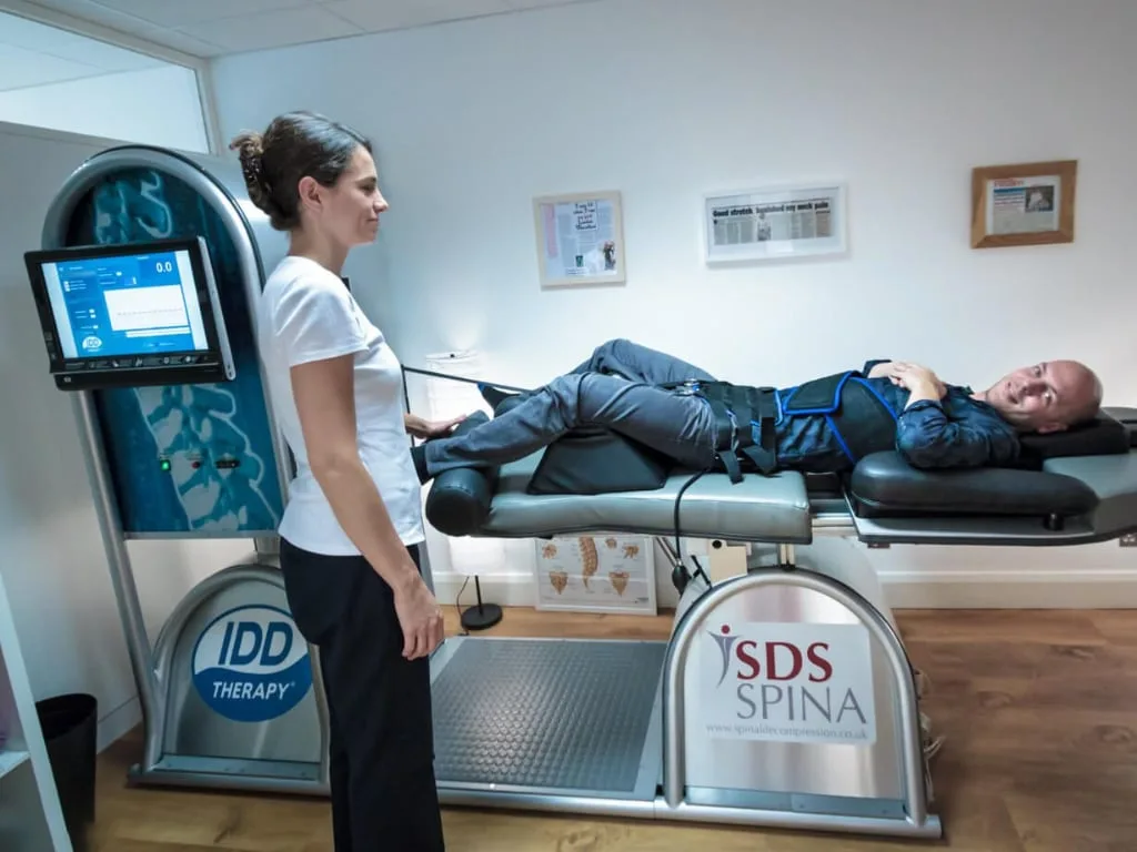
The main treatment we offer at Spine Plus for bulging discs and spondylosis in the neck or lower back is our non-surgical, non-invasive, computerised IDD Spinal Decompression Programme. This involves a course of sessions on our IDD Therapy machine to gently and precisely stretch specific disc segments in order to promote natural disc healing, coupled with a core strengthening exercise regime to improve spinal stabilisation. Where appropriate we often add in other treatment modalities such as paraspinal dry needling (acupuncture) to the deep muscle of the spine to further promote healing and reduce tension on the damaged disc(s). It is important that all potential patients have an initial assessment with one of our clinicians, including a review of their MRI scan(s) to assess their suitability for IDD.
One of the advantages of IDD Therapy is that it is non-invasive and possesses very little risk of harm. However where IDD Therapy is inappropriate or proves not to be effective, we aim to assist clients in deciding on the most appropriate further options for their degenerative disc condition(s).
For information purposes only. The list of injections and surgical procedures below may not be complete, and we ultimately recommend guidance from a spinal surgeon or medical consultant before considering any of them.
Disc & Annular Tears
In order to understand what an annular disc tear is and how it can affect the human body, it is important to first understand what an intervertebral disc is and the essential functions that it is responsible for.
Intervertebral Disc and Annular Disc Tears
The human spine is made up of numerous bones or vertebrae that support the head and torso. Each pair of vertebrae has an intervertebral disc, which serves as a pressurised cushion between them. In all, the human spine has 23 discs that lie in the cervical region, thoracic region and the lumbar region of the spinal column.
An intervertebral disc forms a cartilaginous joint at each vertebra, enabling them to move so that a person can bend and twist. The disc also functions as a fibrous tissue that helps to hold the numerous spinal vertebrae together. Each disc can further be divided into two main parts, namely the annulus fibrosus and the nucleus pulposus.
- Annulus Fibrosus: This is the tough outer capsule of an intervertebral disc and is made up of multiple fibrocartilage that contain a combination of white fibrous tissue as well as cartilaginous tissue. The layers of the annulus have several nerve fibres that are extremely sensitive to pain.
- Nucleus Pulposus: This is a gel-like substance that is contained inside the disc and is protected by the outer covering of the annulus. The nucleus pulposus does not have any nerve fibres supplied to it.
The annulus and the nucleus together make up an intervertebral disc, responsible for absorbing shock and impact and keeping two vertebrae separated from each other. The annulus of a disc is a ligament and as such it is susceptible to tears. When the annulus of a disc gets damaged, it is known as disc and annular tears. When the annulus tears, the soft gel-like substance inside the nucleus can start leaking, leading to a herniated disc. Since the outer layers of the annulus contain several nerve fibres, an annular tear can often be quite painful.
Types of Disc and Annular Tears
Disc and annular tears can be categorised into three main types:
- Rim Lesions: A tear in the outer-most fibres of the annulus, typically horizontal and usually caused by physical trauma. Discs with rim lesions generally degenerate faster than healthy discs.
- Radial Tears: Occurs in the inner-most portion of the disc (the nucleus pulposus), typically from natural ageing, impact or injury. Since these tears occur where there are no nerve fibres, they may be asymptomatic. Graded 0 (healthy) through to 5 (nucleus material seeped through the entire annulus).
- Concentric Tears: A tear between the layers of collagen (lamellae) within the annulus, usually from repetitive physical trauma. Occurs in the outer layers, so can be extremely painful.
Diagnosis is typically done by a test known as discography, where a dye is injected into the disc and the disc is pressurised to determine whether the dye spreads and any pain occurs, though the procedure itself is invasive and can be painful. An alternative diagnostic tool is the HIZ (High Intensity Zone) Sign, a finding on an MRI scan that is a good indicator of a damaged disc.
Causes of Disc and Annular Tears
By the time a person reaches the age of 30, the natural strength of the annulus starts to deteriorate, making it more susceptible to tears from physical trauma. Beyond natural ageing, several factors increase the risk:
- Sudden harsh physical trauma
- In younger individuals with a strong annulus, harsh trauma or sudden impact (a vehicular accident, contact sports) can tear the annulus fibrosus.
- Nature of occupation
- Heavy lifting, pushing, pulling, or sustained strenuous activity can cause the annulus to develop tiny rips that become larger tears over time.
- Being obese
- Excess body weight puts tremendous stress on the spine, compressing intervertebral discs and accelerating degeneration.
- Genetics
- A genetic predisposition to weak annular collagen means even minor physical impact can lead to tears.
Some more risk factors typically associated with disc and annular tears include high impact exercising, extreme sports and excessive smoking.
Symptoms of Disc and Annular Tears
Diagnosing disc and annular tears can be difficult, since a person may not experience any symptoms at all: it depends which layer of the annulus is affected. Only the outer-most layer carries nerve fibres, so a tear there (or one that lets nucleus material reach it) is likely to be painful; a tear confined to the inner or middle layer may cause no symptoms whatsoever. Where symptomatic, the common symptoms are:
- Pain: Depending on which disc is torn, pain is felt in different areas: a torn cervical disc causes neck, shoulder and arm pain; a torn lumbar disc causes lower back, buttock, leg and sometimes foot pain.
- Tingling and numbness: Pins-and-needles sensations and numbness in the arms and legs, sometimes with muscle weakness or spasm, depending on which nerves are affected.
- Swelling: Inflammation of the affected nerves can cause swelling in different parts of the body, again depending on which nerves are impacted.
Most people notice their symptoms and pain worsen with physical activities such as bending, sitting, lifting things, or even sneezing hard, since these movements increase pressure on the damaged disc. Standing, by contrast, can often relieve that pressure.
Treatment and Prevention
Disc and annular tears can be treated with non-surgical options as well as disc tear surgery:
- Non-surgical options: asymptomatic tears often need no treatment at all. Where symptoms occur, the first line is rest and physical therapy, anti-inflammatory drugs for swelling, ice/heat therapy, massage, acupuncture or chiropractic alignment, and core-strengthening exercise (yoga, swimming, brisk walking).
- Surgical treatment: most tears heal on their own via the body's natural scar-tissue process. If non-surgical treatment doesn't reduce the pain, surgery is a last resort, typically one of three types: open back surgery (most invasive, cutting open the skin to reach and remove the torn disc and fusing the affected vertebrae together), laser disc decompression surgery (less invasive, using a laser to shrink the damaged disc), or endoscopic disc tear surgery (minimally invasive, a special surgical tool inserted through a tiny incision to locate and seal the tear).
Prevention is generally simpler than treatment: exercising to enhance core muscle strength and flexibility, maintaining a healthy body weight, and giving up smoking (nicotine accelerates disc degeneration) all reduce the risk of disc and annular tears developing in the first place.
Spinal Injection Therapy
An overview of the interventional injection procedures a spinal specialist may discuss with you. Tap a procedure to read more.
Lumbar Caudal Epidural
A steroid and pain killer are injected into the epidural space (space around the spinal nerve), via the sacral hiatus (small channel at the base of the sacrum just above the buttock cleft).
Cervical Epidural
An injection of a pain killer and anti-inflammatory steroid is inserted into the epidural space of the neck. It is used to ease pain and inflammation often associated with nerve root entrapment and degenerative disc disease.
Nerve Root Block
An injection is performed under x-ray guidance, whereby a pain killer and an anti-inflammatory steroid is injected very near to a specific nerve root within the spine that is transmitting a large amount of pain due to injury or pressure on that nerve root.
(Therapeutic) Facet Joint Injection (steroid)
An injection of a numbing pain killer and anti-inflammatory steroid is inserted into the small facet joints on one or both sides of a vertebral level. The procedure is performed under x-ray guidance and commonly used when the wear and tear of the facet joints is thought to be responsible for a patient's pain. The injections can be used for diagnostic purposes (pain killer only) to determine if a particular facet joint is the source of pain, or for therapeutic purposes to treat the source of the pain. When such injections work, pain relief can be achieved for several months or more, after which the procedure can be repeated. Some clinicians raise concerns that injecting a steroid into synovial joints (including facet joints) may accelerate the rate of degeneration of healthy cartilage within the joint and so may therefore wish to reserve this technique only for patients with severely degenerate joints or if there is a very good chance of pain relief that will outweigh any risks.
(Therapeutic) Sacro-iliac Joint Injection (Steroid)
An injection that is usually performed with live x-ray guidance into one or both sacroiliac joints (joints located at the base of the spine between the sacrum and the pelvic bones). Sacro-iliac joint dysfunction is thought to be the source of low back and buttock pain and can even mimic pain from other structures such as lumbar discs, nerve roots and facet joints. A mixture of a steroid and pain killer is injected into the joint with the intention of providing long term relief and reduction of inflammation within it. Some clinicians raise concerns that injecting steroids into synovial joints (including sacro-iliac joints) may accelerate the rate of degeneration of healthy cartilage within the joint and so may therefore wish to reserve this technique only for patients with severely degenerate joints or if there is a very good chance of pain relief that will outweigh any risks.
Radiofrequency (joint) Denervation
This is a procedure during which the nerves innervating a joint are burnt using a radio frequency probe. This stops pain signals being generated from the joint. The nerve endings eventually grow back, usually after a few months, after which the procedure may be repeated.
Pulsed Radiofrequency Denervation
This is a procedure similar to joint radiofrequency denervation. However, rather than targeting the facet joint, this procedure is usually used to target the nerve root or branches of it. Also, rather than burning all the nerve fibres, pulsed radio waves are used which do not have any heating effect but selectively "stun" the pain-carrying fibres.
Intradiscal Electrothermal or Radiofrequency Modulation
There are a number of procedures available that involve passing heating wires and/or radiofrequency electrodes into the disc with the intention of ablating painful nerve fibres within the disc wall and/or sealing tears in the disc wall.
Intradiscal Chemical Modulation
This procedure involves an injection of drugs, such as corticosteroid or Methyl Blue, into an intervertebral disc with the intention of blocking nerve signals or stabilising the inflammatory process within a painful disc.
Prolotherapy (Proliferation Therapy)
Prolotherapy, sometimes referred to as sclerosing injections, is most commonly undertaken by orthopaedic physicians. A solution containing sugar and phenol is injected into ligaments supporting the spine with the hope of stimulating proliferation of collagen in order to strengthen the ligaments, stabilise the spine and reduce pain.
Spinal Surgery Procedures
Surgical options range widely by technique, region of the spine, and how invasive the approach is. Grouped below by procedure type. Tap a procedure to read more.
Lumbar Fusion
(Posterolateral Gutter) Spinal Fusion
This is generally regarded as the most "tried and tested" method for surgically fusing together two adjacent spinal segments (vertebrae). It involves a three to six inch incision through the midline of the back. A bone graft is harvested from the patient's hip bone (iliac crest) and attached between the "transverse processes" on each side of the vertebrae (such as L4 and L5). Muscles that have been partially dissected during the procedure are then reattached over the graft in order to create tension. Screws, rods and other surgical instrumentation may also be used to encourage a solid fusion by holding the vertebrae in place as the graft heals over time. Whilst the procedure has a long and trusted history, the technique on its own does not offer a way of restoring intervertebral height resulting from a severely thinned disc (spondylosis) and may therefore be reserved for conditions such as sponlylolisthesis, where one vertebrae has slipped forward over another but where the intervertebral disc itself is relatively healthy and does not need to be removed (partially or wholly). The surface area for the graft is smaller and is not exerted to the same amount of body weight compression compared to the "interbody" methods of fusion. This avoids the small risk of graft retropulsion associated with interbody fusion and means the graft does not benefit from compressing forces encouraging bone growth, as opposed to Wolff's law which argues that bones heal better when under compression. This may explain why the chances of achieving solid fusion are reported as being less likely than with the more recently developed interbody methods of fusion.
Posterior Lumbar Interbody Fusion (PLIF)
PLIF is a surgical procedure for fusing together the vertebral bodies of two lower back spinal segments (vertebrae). Most of the damaged disc that lies between the vertebral bodies is removed and replaced with bone graft. This lifts pressure from pinched nerve roots and enables a solid bone mass or "fusion" to form, thus stabilising the spine. With "posterior" interbody fusion, the surgical approach is made posteriorly in the midline of the back. Muscles either side of the spine are stripped away and the laminae and interspinous process from one vertebra are removed so that access can be gained to the intervertebral disc space. The surgeon may also supplement the PLIF procedure with a "Posterolateral Spinal Fusion" whereby a series of screws and rods, as well as bone grafts, are inserted in the back and sides of the vertebrae to provide increased stabilisation.
Anterior Lumbar Interbody Fusion (ALIF)
ALIF is a surgical technique for fusing together the vertebral bodies of two lower back spinal segments (vertebrae). Most of the damaged disc that lies between the vertebral bodies is removed and replaced with bone graft. This lifts pressure from pinched nerve roots and enables a solid bone mass or "fusion" to form, thus stabilising the spine. Surgical access to the disc space is gained from an "anterior" incision through the abdomen. This has the advantage of leaving the bone, muscles and nerves at the back of the spine undisturbed (unlike with other forms of fusion). An anterior approach also allows for a much larger implant, providing better initial stabilisation. However making an incision through the stomach does pose some risk to the important structures in that area, such as major blood vessels, which can require particular expertise from the spinal surgeon and may require an additional (vascular) surgeon to be present during the procedure. For men undergoing an L5/S1 fusion there is also a slight risk (about 1%) of damage to nerve endings controlling valves during ejaculation, meaning that ejaculate can be expelled into the bladder ("retrograde ejaculation"). Erectile function, sensation of ejaculation and orgasm are unchanged, but it can make conception very difficult. For some people a second surgical procedure is planned to take place a week or so after their ALIF surgery to insert screws and/or rods into the back of the spine to provide posterior fixation and additional stabilisation.
Extreme Lateral Interbody Fusion (XLIF)
This is a minimally invasive surgical procedure for fusing together the vertebral bodies of two lower back spinal segments (vertebrae). Access to the intervertebral disc space is gained through a small incision in the patient's side. This allows major muscles of the back to be avoided. Most of the damaged disc that lies between the vertebral bodies is removed and replaced with bone graft. This lifts pressure from pinched nerve roots and enables a solid bone mass or "fusion" to form, thus stabilising the spine. The procedure may be performed on an outpatient basis, however it is not suitable for all conditions where spinal fusion is an option. For example, it cannot be used on the lowest disc space (L5/S1). A particular risk from the lateral approach is the possibility of damage to the lumbar plexus of nerves serving the thigh (quadriceps) muscles. This risk is minimised by use of an electromyograph (EMG) monitor to warn the surgeon if they are near a vulnerable nerve.
Trans-Foraminal Lumbar Interbody Fusion (TLIF)
This is a surgical procedure for fusing together the vertebral bodies of two lower back spinal segments (vertebrae). Most of the damaged disc that lies between the vertebral bodies is removed and replaced with bone graft. This lifts pressure from pinched nerve roots and enables a solid bone mass or "fusion" to form, thus stabilising the spine. With the trans-foraminal approach, the incision is made posteriorly to one side of the midline, with the procedure involving removal of one of the facet joints that connects the two vertebrae together. The main theoretical advantages in comparison to PLIF are that this allows for greater visualisation into the disc space, increased disc removal, a larger implant/bone graft and greater distraction of the disc space. However this must be weighed against the fact that an entire facet joint from one side of the spine is removed (rather than a portion of each facet from either side of the spine as with PLIF).
Posterior Lumbar Dynamic Stabilisation
The instrumentation used during this type of spinal surgery has evolved from the pedicle screws and rods traditionally used during posterior spinal fusion. However rather than using rigid screws and metal rods, flexible screws and rods with moving parts are now used instead. The goal of such surgery is to offer stabilisation of an intervertebral segment without fusion, i.e. allowing controlled motion between the two vertebrae. By preserving near normal movement at the operated level, the hope is to avoid the increased strain on adjacent levels of the spine that can develop several years after traditional fusion operations.
Lumbar Decompression & Minimally Invasive
Lumbar Decompression (Laminectomy)
The lamina is the name of part of a vertebral segment that forms a bony ring or "canal" around the spinal cord and spinal nerves. This canal becomes narrowed in conditions such as spinal stenosis, causing encroachment on the spinal cord and nerves. Spinal stenosis most commonly affects older age groups who suffer generalised wear and tear in the spine. Lumbar spinal stenosis affects the lower back and usually manifests as leg pain whilst walking. "Laminectomy" surgery was developed in the early 1900s and involves a three to five inch incision being made in the lower back. The paraspinal muscles are dissected off the lamina on both sides of the spine, the lamina and spinous process are then completely removed, effectively making the spinal canal at that segment into a semicircle rather than a ring of bone, which frees up or "decompresses" the nerves. Success rates for relieving leg symptoms from spinal stenosis is around 70-80%, although a laminectomy is less reliable for relieving back pain. There is around 1–3% chance of complications occurring, such as nerve damage, incontinence and infections. Also there is a risk for some patients that by removing many of the stabilising muscles and ligaments of the vertebra, segments of the spine may become destabilised, leading to chronic back pain and weakness. Stenosis symptoms can also reoccur several years after the surgery, when the degenerative processes that originally caused the stenosis continue. In some cases where stenosis is combined with an unstable segment, stenosis may be prevented from reoccurring by performing a fusion at the same time as the laminectomy. For some people, an alternative to laminectomy is laminoplasty, which involves a smaller incision, removing the lamina from only one side of the vertebra, thus preserving more of the supporting structures of the spine.
Lumbar Laminoplasty
A posterior surgical technique developed in the 1980s as a less invasive alternative to lumbar laminectomy, which involves cutting through the lamina on just one side of a vertebra (rather than both sides as in a laminectomy). This creates a hinge in the lamina which is used to widen the spinal canal in order to decompress the nerves. The newly widened canal is then sealed with a bone graft. One of the main advantages of laminoplasty is that, in contrast to a laminectomy, much of the supporting structures of the vertebra, such as the muscles, ligaments and spinous process, are preserved. Despite the fact that laminoplasty is a less invasive alternative to laminectomy, some surgeons do not offer it because it is a more time-consuming operation and requires specialised training and equipment, such as an operating microscope.
Lumbar Micro-Discectomy
This is one of the most widely available minimally invasive surgical techniques for treating sciatica due to a pinched nerve root from a herniated or bulging disc. The surgeon uses an operating microscope or glasses (Loupes) in order to reduce the size of the incision needed to access the disc. A small 1 to 2 inch incision is made, slightly to the side of the midline in the lower back. Muscle tissue is gently divided and a small amount of ligament and bone is removed from the lamina and/or facet joint in order to create a window through which the surgeon can remove the bulging portion of the disc and free the trapped nerve root. Lumbar microdiscectomy is reportedly 95% successful at reducing sciatic pain in the leg, with the best outcomes experienced by patients who have had sciatica for less than three to six months. Many cases of sciatica caused by an isolated disc bulge will improve on their own or with the help of physical therapy. If such improvement is going to be achieved without surgery, it will generally happen within 6 to 12 weeks. Therefore, except for severe cases or in the presence of "Cauda Equina Syndrome", the usual advice is to wait at least 6 weeks before considering surgery. There is a small risk (5–10% chance) of the disc bulge reoccurring after surgery, which is more likely within the first 3 months, but can occur several years after surgery. For this reason some surgeons excavate some of the inner part of the disc in addition to removing the bulging portion so that there is less disc material capable of herniating again in the future. This must be weighed against the fact that the more of the disc material removed, the less of it there will be to perform its roles of providing shock absorption and height between the vertebrae and exiting nerve roots. Excessive disc excavation could potentially lead to premature load and degeneration on other structures of the spine, such as the facet joints, several years later.
Endoscopic (Keyhole) Lumbar Discectomy
This is a minimally invasive surgery performed through a small diameter tubular device inserted into the back. This technique is most commonly performed to remove the herniated or prolapsed portion of an intervertebral disc that is compressing the adjacent nerve root causing sciatic pain in the leg. Unless using a "transforaminal approach", access to the spinal nerves is gained through the midline, which involves removing small amounts of bone, similar to a microdiscectomy. An endoscopic lumbar discectomy is designed to be even more minimally invasive than using a microscope (microdiscectomy). Popularity for endoscopic lumbar discectomy is lacking amongst some surgeons because of concerns it gives limited visualisation of the disc compared to microdiscectomy. The complication rate is also reported as being higher, with the procedure requiring specialised training and being technically more difficult to perform.
Transforaminal Endoscopic Lumbar Discectomy
This is keyhole lower back surgery, whereby an endoscope is used to "walk up the nerve" through the neural foramen (the hole through which the nerve root emerges from the bony spinal canal). This "transforaminal" approach is designed to cause the least amount of disruption to the spinal musculature and bony architecture (even less than traditional endoscopic spinal surgery), whilst at the same time allowing for good visualisation. Once in the foramen, the bulging portion of a herniated disc can then be removed in order to reduce nerve root compression.
Trans-Foraminal Endoscopic Lumbar Decompression & Foraminoplasty (TELDF)
As above, this is keyhole lower back surgery, whereby an endoscope is used to "walk up the nerve" through the neural foramen (the hole through which the nerve root emerges from the bony spinal canal). This "transforaminal" approach is designed to cause the least amount of disruption to the spinal musculature and bony architecture whilst at the same time allowing for good visualisation of the nerve, disc, facet and associated ligaments. Once in the foramen, as well as removing bulging portions of intervertebral discs, the neural foramen can be cleared of scar tissue, nerve adhesion and bone spikes with the use of a laser. This type of surgery is not widely available and is technically difficult, requiring specialised equipment and training.
(Automated) Percutaneous Lumbar Discectomy
This is the most minimally invasive of all the surgical techniques, designed to reduce an intervertebral disc protrusion. The procedure is performed under x-ray guidance using a probe or needle inserted into the affected disc. The intention is that there is the least amount of disruption to the posterior spinal muscles and other soft tissues. In reality, the surgeon cannot visualise or access as much of the disc as with other minimally invasive procedures, such as microdiscectomy, and the success rates are not as good, however it may be appropriate for some selected cases. If the procedure fails the patient may be able to move on to others, such as microdiscectomy or endoscopic surgery.
Percutaneous Nucleoplasty
This procedure is usually performed on an outpatient basis, involving a device introduced via a hollow needle that removes part of the inner nucleus of an intervertebral disc in order to reduce pressure within it, thereby shrinking the disc so that it exerts less pressure on neighbouring structures, such as pinched nerves.
Devices & Disc Replacement
Facet Replacement or Total Element Replacement Devices for Spinal Stenosis
These relatively new surgical implants are designed to replace the facet joints and associated bony projections that form the posterior architecture of a vertebral segment in the spine. They are primarily used for patients with unresolved facet joint pain and/or spinal stenosis caused by severe degeneration of the facet joints. These devices are designed to preserve movement of the spinal segment, unlike spinal fusion, yet allowing more stabilisation and control of movement than a total laminectomy. However these devices are relatively new on the market and do not have the outcome history of more extensively used procedures.
Lumbar Disc Replacement
Offered by some surgeons as an alternative to lumbar fusion, whereby a damaged intervertebral disc (the cartilaginous cushion that sits between two vertebrae) is replaced with an artificial one. Advantages over spinal fusion are that the surgery is less complex and is quicker to perform, with the main theoretical advantage being that movement between the vertebrae is preserved, thus reducing the negative effects on adjacent levels that can occur with spinal fusion. More recent styles of prosthetic discs also offer shock-absorbing qualities. Lumbar artificial disc surgery has been available in European countries for around 15 years. Currently only around 50% of spinal surgeons offer the procedure, often because they are sceptical about the reality of how much movement the prosthesis allows or are concerned about the possibility of the artificial disc becoming dislodged.
Lumbar Interspinous Distraction Devices (e.g. X-Stop)
A small prosthetic spacing device is inserted between the spinous process of two adjacent vertebrae to limit the amount of backward bending movement (extension) allowed at that level of the spine, thus preserving the patency of the foramina or tunnels through which the spinal nerves pass. This procedure is designed to alleviate pressure on spinal nerves experienced in conditions such as spinal stenosis. Some reports have also suggested that, in some cases, patients will experience other unintended benefits, such as reduced pressure on painful degenerative discs. The procedure is relatively inexpensive and since the spinous processes lie near the surface of the body, the procedure is relatively uncomplicated and may be performed on an outpatient basis. However it is by no means suitable for everyone: for example, people with weakened bones, such as those with osteoporosis, may be at risk of the device causing a fracture to their spinous processes.
Cervical (Neck) Procedures
Anterior Cervical Discectomy & Fusion (ACDF)
ACDF is one of the standard techniques used to treat degenerative disc disease in the neck (cervical spine). An incision is made through the front of the neck to avoid the delicate nerve endings and spinal cord that would be vulnerable from accessing the spine with a posterior incision. The damaged disc and debris is removed, ready for insertion of a spacer device containing a bone graft. This lifts pressure off of nerve roots and the spinal cord, which may have been caused by the damaged disc. The vertebrae above and below the disc space may be fixed together using a plate and screws. Over time the bone graft knits the two vertebrae together, forming a solid stable bone mass or "fusion". This is a reliable technique with good success rates and outcomes. However there are concerns that, for some individuals in the longer term, fusing two vertebrae together, thereby eliminating movement at that level, may cause premature wear and tear at other nearby levels of the spine.
Anterior Cervical Corpectomy
This surgery is designed to treat severe cervical stenosis or diseased vertebral bodies in more than one level of the neck. This procedure is completed through an incision in the front of the neck. The surgeon removes one or more vertebral bodies as well as discs above and below each segment in order to fully decompress the spinal canal. The defect is then reconstructed with a bone graft to create a multilevel fusion. This is extensive surgery performed on cases involving significant deformity, therefore the risks of complications, such as damage to the spinal cord and nerves, are higher than with other procedures.
Cervical Laminectomy
This type of surgery is completed through a 3 to 4 inch incision in the back of the neck. During the procedure the lamina, spinous process and overlying muscle on both sides of the neck is removed from one segment of the cervical spine. This effectively turns the ring of bone called the spinal canal, through which the spinal cord passes, into a semicircle. This alleviates pressure on the spinal cord in cases where it was being compressed due to narrowing of the bony spinal canal. The main risk from this operation is that damage could be caused to the spinal cord, nerves and membranes. To lessen this risk, the surgeon will often use specialised electrodes to monitor spinal cord function during the procedure (Somatosensory Evoked Potentials – SSEPs). By cutting away much of the supporting muscle, ligament and bone of the operated segment there is also risk of the spine at that level developing instability and deformity. Therefore cervical laminectomy is sometimes performed in combination with cervical fusion surgery.
Cervical Laminoplasty
This is a surgical technique involving cutting through one side of part of a bony segment in the neck called the lamina. The lamina forms part of a ring of bone at each vertebra, called the spinal canal, through which the spinal cord passes. This canal becomes narrowed in conditions such as spinal stenosis, causing encroachment of the spinal cord and nerves. Once an opening has been created in the lamina, this creates a hinge allowing the spinal canal to be widened, with a bone graft then used to seal the opening. An attractive feature of cervical laminoplasty is that, unlike fusion, it preserves movement of the operated segment, therefore lessening the likelihood of premature degeneration of adjacent segments. Also, compared to laminectomy, much of the muscle, ligament and bone is preserved, thus reducing the likelihood of post-operative instability. However by approaching the cervical spine posteriorly and cutting through the lamina on just one side, it may be more difficult for the surgeon to assess whether or not the canal has been well decompressed. Also, there is a small amount of risk of damage to the spinal cord and nerves exposed in the area during such a posterior approach. This risk is less for other procedures used for spinal stenosis that access the spine from the front of the neck, such as Anterior Cervical Discectomy and Fusion (ACDF).
Cervical Disc Replacement
This is the surgical replacement of a damaged or degenerated disc in the neck, initiated by making an incision in the front of the neck. This enables the surgeon to avoid the delicate nerve endings and spinal cord that would be vulnerable from accessing the spine posteriorly. The defective disc is removed, the area cleaned and a new artificial disc is inserted into the disc space. This lifts pressure off of nerve roots and the spinal cord at the same time as maintaining movement at that level of the spine. Artificial disc replacement in the neck is now an established reliable technique, although it is not as "tried and tested" as other more traditional techniques, such as Anterior Cervical Discectomy and Fusion (ACDF). The obvious advantage over ACDF is that it allows movement of the vertebral segment and so reduces the chance of premature adjacent level wear and tear that is associated with fusions.
Cervical Endoscopic Spinal Surgery
This keyhole surgery performed on the cervical spine (neck) is most commonly performed to decompress trapped nerves caused by disc herniation. The advent of such technology in the cervical spine has allowed for small incisions and less disruption to muscle and other tissues than with other more invasive techniques, such as disc fusion or disc replacement. Also, whereas removal of the whole disc may once have been a patient's only option, where appropriate, cervical endoscopic spinal surgery potentially means removing just the herniated portion of a cervical disc is possible whilst preserving its remaining healthy portion. Popularity for endoscopic spinal surgery is lacking amongst some surgeons because of concerns it gives limited visualisation, the procedure requires specialised training and is technically more difficult to perform.
Thoracic (Mid-Back) Procedures
Thoracic Laminectomy (Thoracic Spine Surgery)
Spinal surgery is performed on the middle back (thoracic spine), which is the part of the spine where the rib cage attaches to it. Thoracic spine surgery is far less common than neck or lower back surgery because degenerative changes in the discs and joints are often asymptomatic. It is usually only reserved for rare cases that cause spinal cord compression, progressive neurological deficit and intolerable pain. Thoracic laminectomy is the traditional historical approach to thoracic spine surgery, involving an approach through the midline of the spine, removing the lamina (portion of a vertebral bone) and pushing the spinal cord aside in order to gain access to degenerative discs. This approach has now largely been replaced by more minimally invasive procedures.
Thoracic Costo-Transverse Ectomy (Thoracic Spinal Surgery)
Spinal surgery is performed on the middle back (thoracic spine), which is the part of the spine where the rib cage attaches to it. Thoracic spine surgery is far less common than neck or lower back surgery because degenerative changes in the discs and joints are often asymptomatic. It is usually only reserved for rare cases that cause spinal cord compression, progressive neurological deficit and intolerable pain. During the Costo-Transverse Ectomy procedure, a small amount of vertebral bone (transverse process) and rib is removed in order to gain access to the thoracic spine.
Anterior Trans-Thoracic Surgery (Thoracic Spine Surgery)
Spinal surgery is performed on the middle back (thoracic spine), which is the part of the spine where the rib cage attaches to it. Thoracic spine surgery is far less common than neck or lower back surgery because degenerative changes in the discs and joints are often asymptomatic. It is usually only reserved for rare cases that cause spinal cord compression, progressive neurological deficit and intolerable pain. Trans-thoracic surgery involves approaching the thoracic spine through the front, either via an open procedure through the chest cavity or, in some centres, via a minimally invasive procedure involving a number of small incisions, arthroscopes and a video screen (Video Assisted Thoracic Surgery, VATS).
Video Assisted Thoracic (Spine) Surgery (VATS)
This minimally invasive surgery for the middle back (thoracic spine) involves an anterior approach through the chest cavity, using a number of small incisions, arthroscopes and a video screen.
Other
Awake State Spinal Surgery
Spinal surgery is performed using an endoscope whilst the patient is sedated, but conscious, to enable the patient to provide instant feedback to the surgeon about sensitive pain-provoking structures. This technique is most appropriate in cases where there may be some doubt as to the exact cause of a patient's pain pattern, so that the source of pain can be more accurately identified, rather than relying on what "looks" likely from an MRI scan.
Call us on 020 8506 1000 to discuss your disc problem and treatment options.